
In G/GEJ tumours, CLDN18.2 is reported as the percentage of tumour cells stained with moderate-to-strong membranous stain intensity.1
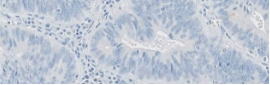
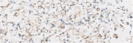
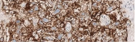
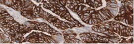
- Tissue slides with tumour cells present can demonstrate varying levels of CLDN18 membranous staining intensity, ranging from no staining to strong staining (0 to 3+)
- Many studies have included percentage of tumour cells with only moderate-to-strong (2+/3+) membranous staining for scoring CLDN18
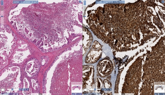
Account for tumor necrosis during interpretation.
Click to view stain gallery

References: 1. Pellino A, Brignola S, Riello E, et al. Association of CLDN18 protein expression with clinicopathological features and prognosis in advanced gastric and gastroesophageal junction adenocarcinomas. J Pers Med (Epub) 10-26-2021. 2.Rohde C, Yamaguchi R, Mukhina S, et al. Comparison of Claudin 18.2 expression in primary tumors and lymph node metastases in Japanese patients with gastric adenocarcinoma. Jpn J Clin Oncol 2019;49(9):870-6.